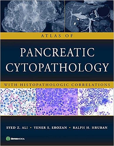
Download Atlas of Pancreatic Cytopathology with Histopathologic by Syed Z. Ali MD, Yener S. Erozan MD, Ralph H. Hruban MD PDF

By Syed Z. Ali MD, Yener S. Erozan MD, Ralph H. Hruban MD
Scientific and radiologic examinations can't reliably distinguish benign or inflammatory pancreatic sickness from carcinoma. The elevated use of pancreatic tremendous needle aspiration (FNA) in addition to advances in imaging strategies and the creation of endoscopic ultrasound counsel have ended in a lot better detection and popularity of pancreatic plenty. accordingly, pancreatic cytopathology is vital to actual pre-operative analysis, but it's a hard diagnostic zone with various capability pitfalls and ???„????look-alike???„???? lesions. Skillful popularity and an expertise of the restrictions of the approach are crucial in averting misdiagnosis of those harmful lesions. Atlas of Pancreatic Cytopathology with Histopathologic Correlations fills a void in present pathology literature. With 450 high-resolution photos, together with photos of histopathologic and radiologic beneficial properties, this useful atlas offers an built-in method of diagnostic cytopathology that might support health care professional cytopathologists, cytotechnologists, and pathologists stay away from power pitfalls and ""look-alike"" lesions. Written through well-known specialists within the box, the broad high-resolution colour photographs of the attribute beneficial properties of pancreatic affliction are offered with special descriptions that disguise vintage positive factors, diagnostic clues, and power pitfalls. Atlas of Pancreatic Cytopathology with Histopathologic Correlations is a invaluable source for the professional cytopathologist, basic and surgical pathologists, pathology trainees, and cytotechnologists.
Read or Download Atlas of Pancreatic Cytopathology with Histopathologic Correlations PDF
Similar digestive organs books
Principles and Practice of Gastrointestinal Oncology
Completely up-to-date for its moment version, this article offers entire, interdisciplinary insurance of gastrointestinal melanoma, together with molecular biology, analysis, clinical, surgical, and radiation treatment, and palliative care. The preliminary part, ideas of Gastrointestinal Oncology, comprises an improved radiation oncology bankruptcy, an greatly revised melanoma genetics bankruptcy, and a very rewritten scientific oncology bankruptcy emphasizing new brokers.
This can be a 3-in-1 reference ebook. It offers an entire scientific dictionary overlaying hundreds and hundreds of phrases and expressions on the subject of hiatus hernia. It additionally offers huge lists of bibliographic citations. eventually, it presents info to clients on the right way to replace their wisdom utilizing a variety of web assets.
It really is with a lot excitement that I introduce this primary quantity in a sequence of themes in Gastroenterology geared toward the clever clinician. Dr. Peter Banks is in the beginning a clinician and instructor and accordingly a great lead-off writer. His very useful evaluation of pancreatitis relies not just on a radical assimilation of scientific and experimental proof but additionally on his lengthy scientific perform in college hospitals and in deepest perform.
- Rifaximin: A Poorly Absorbed Antibiotic. Pharmacology and Clinical Use
- Hepatocellular Carcinoma and Liver Metastases: Diagnosis and Treatment
- Endosonography in Gastroenterology
- Lactose Intolerance - A Medical Dictionary, Bibliography, and Annotated Research Guide to Internet References
- Hepatology at a Glance
Extra resources for Atlas of Pancreatic Cytopathology with Histopathologic Correlations
Sample text
They have disorganized and enlarged nuclei, which appear hyperchromatic, forming three-dimensional structures. The changes are not enough to justify an “atypical” or “suspicious” interpretation. 11 — Chronic pancreatitis. Numerous fragments of glandular epithelium, fibrous tissue, and marked background chronic inflammation give this smear a hypercellular appearance. The polymorphous cellular composition clearly favors a benign reactive process in this case. 12 — Chronic pancreatitis. Two fragments of “atypical” columnar epithelium are present.
The mass is very subtle, and the main clue that a tumor is present is the dilatation of the pancreatic duct. (B) Coronal reconstruction of contrast-enhanced CT with maximum intensity projection in the venous phase highlights focal narrowing of the portal confluence (arrow), indicating the tumor has encased the portal confluence and is not resectable. Chapter 2: Radiologic Characteristics of Pancreatic Disease 19 Selected Cases Illustrating Salient Radiologic Characteristics Case 4 Adenocarcinoma of the pancreas unresectable due to arterial and venous encasement (61-year-old man with right upper quadrant and back pain).
Despite the lack of cellular material, this picture strongly suggests chronic pancreatitis with associated fat necrosis. 5 — Chronic pancreatitis. Inflammatory cells with predominance of lymphocytes, display nuclear crush artifact. The cytopathologic appearance is non-specific and chronic pancreatitis is often a diagnosis of exclusion. 6 — Chronic pancreatitis. A large disorganized mesenchymal tissue fragment signifies extensive fibrosis that accompanies such cases resulting in a relatively lack (or absence) of pancreatic epithelium.


