Download Gastrointestinal Cancer Atlas for Endoscopic Therapy by Rikiya Fujita, Hiroshi Takahashi PDF
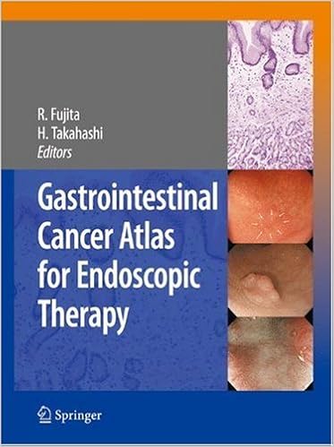
By Rikiya Fujita, Hiroshi Takahashi
Since a couple of mucosal digestive tract melanoma detected has elevated in recent times with an enhance of endoscopic tools, endoscopic remedy of digestive tract melanoma is extensively unfold in Japan yet no longer in different international locations. Minute cancers appear to have personal attribute mucosal indicators in comparison with benign lesions. consequently, ideas, these are various in Japan from different nations, of endoscopic analysis are very important.
The melanoma institute medical institution of JFCR (Japan beginning of melanoma examine) is likely one of the best associations which do the easiest endoscopic therapy in Japan. This e-book is anticipated to be the 1st beneficial atlas within which the considerable studies of this sanatorium supply attractive images of minute cancers with none scars of biopsies which swap facets of unique lesions. This ebook additionally provide photos of magnifying endoscopy utilizing NBI (Narrow Band Imaging) and pathological findings.
Read or Download Gastrointestinal Cancer Atlas for Endoscopic Therapy PDF
Best digestive organs books

Principles and Practice of Gastrointestinal Oncology
Completely up-to-date for its moment variation, this article presents complete, interdisciplinary assurance of gastrointestinal melanoma, together with molecular biology, prognosis, clinical, surgical, and radiation remedy, and palliative care. The preliminary part, ideas of Gastrointestinal Oncology, comprises an elevated radiation oncology bankruptcy, an largely revised melanoma genetics bankruptcy, and a totally rewritten scientific oncology bankruptcy emphasizing new brokers.


This can be a 3-in-1 reference e-book. It supplies an entire scientific dictionary masking countless numbers of phrases and expressions with regards to hiatus hernia. It additionally supplies vast lists of bibliographic citations. ultimately, it offers info to clients on tips on how to replace their wisdom utilizing a variety of web assets.
It truly is with a lot excitement that I introduce this primary quantity in a chain of subject matters in Gastroenterology geared toward the clever clinician. Dr. Peter Banks is at the beginning a clinician and instructor and consequently an excellent lead-off writer. His very worthy evaluation of pancreatitis is predicated not just on an intensive assimilation of scientific and experimental proof but in addition on his lengthy medical perform in collage hospitals and in deepest perform.
- Hepatocellular Carcinoma: A Practical Approach
- Management of Gastrointestinal Diseases in the Elderly
- Gilbert's Syndrome: A Medical Dictionary, Bibliography, And Annotated Research Guide To Internet References
- Living with Crohn's & Colitis Cookbook: Nutritional Guidance, Meal Plans, and Over 100 Recipes for Improved Health and Wellness
- Managing Gastrointestinal Complications of Diabetes
Extra resources for Gastrointestinal Cancer Atlas for Endoscopic Therapy
Example text
Signet-ring cell carcinoma (sig) was diagnosed by biopsy c After marking at ESD. After marking a distance 5 mm from the margin of the lesion, ESD was carried out ous lesion can potentially be removed by biopsy, it is necessary to resect a wider area in one piece to allow detailed pathological evaluation. Such a lesion is a good candidate for ESD. , a biopsy scar). In this respect, ESD is more useful than EMR because the specimen obtained by ESD sustains less mechanical injury than that obtained by EMR (Refer to case 22, Early Gastric Cancer).
Endoscopy 36:1080–1084 Endoscopic Treatment of Minute Gastric Cancer in Cases of a Disappearing-Cancer Biopsy Yorimasa Yamamoto Introduction When diagnosing early gastric cancer by endoscopy, a lesion diameter of 3 mm has been thought to represent the lower limit of detection (1). In general, minute cancer is defined as a tumor ≤5 mm in diameter. Owing to the advances in endoscopic devices, it has become possible to diagnose gastric cancer preoperatively using magnifying endoscopy with narrow band imaging (NBI) (2), although a pathological diagnosis by biopsy is usually employed.
In addition, marginal swelling was found in 36 cases (42%). In contrast, all undifferentiated cancer cases (14 cases) had a pale brownish color, with both encroachment and marginal swelling being found in only 3 cases. Regarding the diagnosis of 0-IIc early gastric cancer, lesions accompanied by marked encroachment were easy to diagnose qualitatively. However, in minute gastric cancers, malignant findings such as “abrupt ending of the fold,” “encroachment of the border,” and “irregularity of the depressed surface” were rarely observed, making qualitative endoscopic diagnosis difficult.