Download Introduction to Functional Magnetic Resonance Imaging: by Richard B. Buxton PDF

By Richard B. Buxton
Read or Download Introduction to Functional Magnetic Resonance Imaging: Principles and Techniques,Second Edition PDF
Best electricity and magnetism books
Magnetic Reconnection: MHD Theory and Applications
Magnetic reconnection is on the center of many dynamic phenomena within the universe, resembling sunlight flares, geomagnetic substorms and tokamak disruptions. Written via global leaders at the topic, this quantity offers a finished review of this primary technique. assurance offers either a pedagogical account of the fundamental conception and a wide-ranging assessment of the actual phenomena created via reconnection--from laboratory machines, the Earth's magnetosphere, and the Sun's surroundings to flare stars and astrophysical accretion disks.
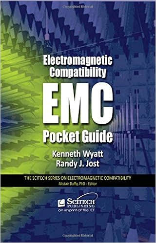
Electromagnetic Compatibility Pocket Guide: Key EMC Facts, Equations, and Data
Each electrical product designed and synthetic world wide needs to meet electromagnetic compatibility (EMC) rules. while you're a operating engineer or technician, the Electromagnetic Compatibility Pocket consultant: Key EMC proof, Equation and information is your quickest and simplest route to the solutions you must in attaining compliance on your designs.
- Plasma Transport in stochastic magnetic field
- Electromagnetic Theory for Electromagnetic Compatibility Engineers
- Hydromagnetic Waves in the Magnetosphere and the Ionosphere
- Electromagnetic Rotary Engine - US Patent 03890548
Additional resources for Introduction to Functional Magnetic Resonance Imaging: Principles and Techniques,Second Edition
Example text
The major amplification comes when a neurotransmitter binds to a post-synaptic receptor and opens a Na+ channel, which may let a thousand ions pass through before it closes again. In this way, the weak signal associated with one neurotransmitter molecule binding to a receptor is amplified a thousand-fold. But all that Na+ must be pumped back in the recovery stage. Very roughly, moving any molecule against its gradient requires about one ATP, so it is perhaps not so surprising that the primary signal amplification stage is the dominant energy cost in neural signaling.
The free energy to drive this uphill pumping is provided by the NADH/NAD+ system, by the transfer of electrons from NADH to the complexes, leaving NAD+. These complexes are arranged in a chain, and the electrons are passed along the chain. At each step in the complex this electron transfer is a thermodynamically downhill process that is coupled to the uphill process of pumping H+ across the membrane against its gradient. At the end of the electron transfer chain, the electron reaches an enzyme called cytochrome oxidase, and the final step in this process is the transfer of four electrons from cytochrome oxidase to an O2 molecule to form two molecules of water.
Such time–activity curves can be analyzed with a kinetic model to extract estimates of individual rate constants for uptake of glucose from the blood and for the first stage of glycolysis. However, the power of the technique is that the distribution of the tracer at a late time point directly reflects the local glucose metabolic rate. In order to derive a quantitative measure of glucose metabolism with either the DG or FDG technique, two other quantities are required. The first is a record of the concentration of the tracer in arterial blood from injection up to the time of the PET image (or the time of sacrifice of the animal in an autoradiographic study).


